Malaria diagnosis has remained largely unchanged for decades, but a team of researchers from the Singapore-MIT Alliance for Research and Technology (SMART) is transforming the game. They’ve harnessed Magnetic Resonance Relaxometry (MRR), akin to MRI, to detect a crucial waste product in the blood of malaria-infected individuals. This revolutionary approach promises a more reliable diagnosis with minimal room for human error.
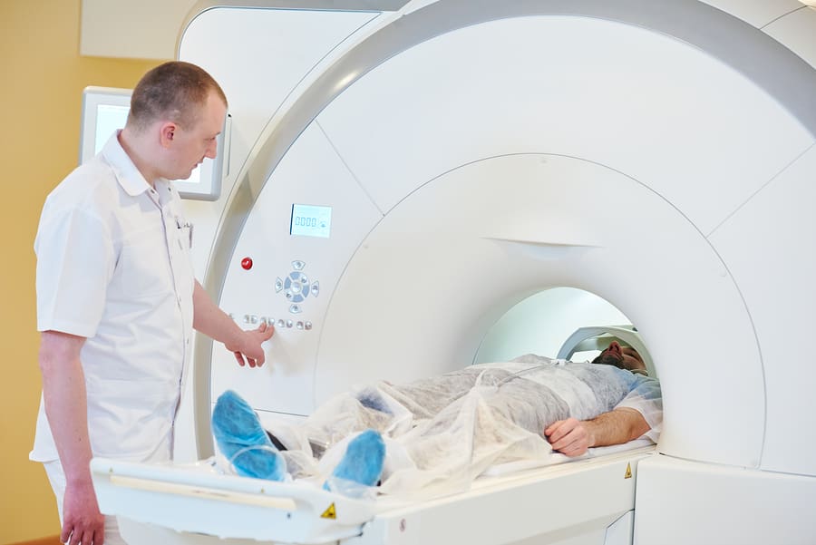
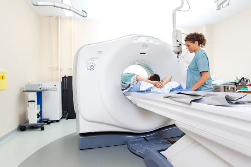
MRI and MRR Scans
MRI (Magnetic Resonance Imaging) and MRR (Magnetic Resonance Relaxometry) scans are related imaging techniques that share some common principles but have distinct differences:
Purpose:
MRI: MRI is a versatile imaging technique primarily used to visualize the internal structures of the body, including organs, tissues, and the brain. It provides detailed anatomical images and is commonly used for diagnosing a wide range of medical conditions.
MRR: MRR, on the other hand, is a specialized application of MRI technology with a specific focus on detecting and quantifying specific molecular components or biomarkers within the body. It is used for highly targeted diagnostic purposes, often related to specific diseases or conditions.
Detection Method:
MRI: MRI primarily relies on the detection of hydrogen protons in water and fat molecules in the body. It captures the response of these protons to a strong magnetic field and radiofrequency pulses to create detailed anatomical images.
MRR: MRR, similar to MRI, uses magnetic resonance principles but focuses on the relaxation times of specific molecules. It measures the time it takes for the magnetic spins of targeted molecules to return to their normal state after being perturbed by radiofrequency pulses.
Information Obtained:
MRI: MRI provides detailed structural information, allowing clinicians to visualize organs, tissues, and abnormalities like tumors, injuries, or inflammation.
MRR: MRR provides information about the concentration and characteristics of specific molecules or biomarkers in the body. It can be used to detect and quantify substances related to certain diseases or conditions, making it highly specific for diagnostic purposes.
Applications:
MRI: MRI is widely used in various medical fields, including neurology, cardiology, oncology, and musculoskeletal imaging. It helps diagnose and monitor a broad spectrum of health issues.
MRR: MRR has specialized applications, such as detecting parasitic waste products in malaria-infected blood or quantifying specific molecules associated with particular diseases. It is not as commonly employed as standard MRI.
Early Malaria Detection and Enhanced Accuracy
The SMART system’s core feature lies in its capability to identify hemozoin, a byproduct generated by malaria parasites. These parasites invade red blood cells and consume hemoglobin, leading to the formation of hemozoin crystals at all stages of malaria infection. This breakthrough technique outperforms traditional methods in sensitivity, accuracy, and efficiency, requiring only a small blood sample. What’s truly remarkable is its simplicity—no special labels or dyes are necessary, thanks to a naturally occurring biomarker.
MRI Sheds Light on Brain Responses to Malaria
Recent research utilizing MRI technology has uncovered intriguing insights into how malaria impacts the brain. A study focused on returning travelers with non-cerebral malaria detected MRI abnormalities, including clinically silent splenial lesions with occasional diffusion restriction. These lesions, previously observed in children with cerebral malaria and uncomplicated endemic malaria, hint at a broader connection to disease severity.
Early Detection for Better Outcomes
While a direct correlation between parasitaemia levels and splenial lesions was not established, these MRI abnormalities may hold promise as prognostic indicators. Detecting these brain imaging findings early could help predict the development of complicated or cerebral malaria. However, further research with a larger sample size and follow-up is essential to solidify this hypothesis.
A Glimpse into Brain Microbleeds in Malaria Patients
The study also employed susceptibility-weighted imaging (SWI) to identify brain microhaemorrhages in returning travelers with non-cerebral malaria. In contrast to prior research in children with cerebral malaria, this study found a low frequency of microhaemorrhages, potentially aligning with the general population’s expectations. Surprisingly, these microhaemorrhages did not show any correlation with parasitaemia levels or clinical symptoms.
White Matter Abnormalities – A Microvascular Mystery
Most patients in the study displayed T2-hyperintense foci of white matter signal abnormalities, suggesting a microvascular origin. Although these abnormalities did not correlate with parasitaemia levels, their link to malarial infection remains uncertain. Further research is needed to unravel the connection between these brain signal changes and malaria.
Unlocking the Potential of MRI in Malaria Diagnosis and Research
MRI technology is not only transforming malaria diagnosis but also offering valuable insights into its impact on the brain. The SMART system’s potential to provide a more reliable and accessible diagnostic method holds great promise for enhancing malaria management globally. As research progresses, we anticipate that MRI technology and MRI Technologists will continue to play a vital role in our fight against this deadly disease.