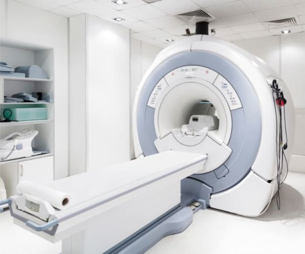
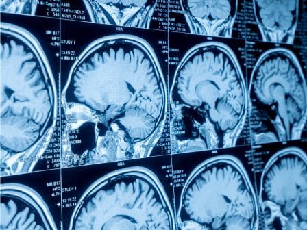
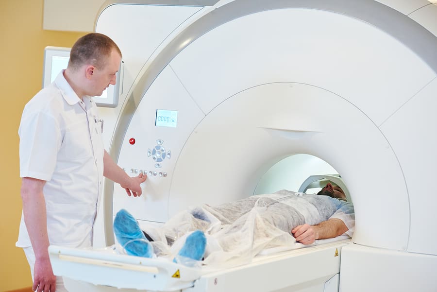
MRI technicians are healthcare professionals specialized in using Magnetic Resonance Imaging (MRI) equipment. This advanced technology allows them to capture detailed images of the inside of the human body. The images produced by MRI machines are crucial for doctors to diagnose and treat various medical conditions accurately. Unlike X-rays, MRIs use powerful magnets and radio waves to create clear pictures of organs, tissues, and skeletal structures without exposing patients to radiation. This makes MRI an invaluable tool in modern medicine, providing insights into the body’s inner workings in a safe and non-invasive way.
The Importance of Effective Communication
In the world of healthcare, the way information is shared is just as crucial as the treatments provided. Effective communication is the backbone of high-quality care. For MRI technicians, this means not only mastering complex machinery but also ensuring that they can convey vital information clearly to both patients and medical teams. Good communication helps in making patients feel at ease, clarifying their doubts, and preparing them for what to expect during the MRI procedure. It bridges the gap between technical expertise and patient care, creating a supportive environment that enhances the overall healthcare experience.
Moreover, MRI technicians collaborate closely with doctors, nurses, and other healthcare professionals. Sharing accurate and timely information about a patient’s condition is essential for diagnosing and deciding on the best treatment plans. In essence, effective communication can significantly impact patient outcomes, making it a critical skill in the fast-paced, high-stakes environment of medical diagnostics.
MRI Terminology: A Closer Look
Navigating the world of MRI technology involves understanding a unique set of terms and phrases. Here’s a closer look at some key MRI terminology, demystified for clarity:
Magnetic Resonance Imaging (MRI): This cutting-edge tool allows us to see inside the human body in detail without surgery. Using a combination of strong magnetic fields and radio waves, MRI machines create images that help doctors diagnose and monitor medical conditions.
Tesla (T): This is the unit of measurement for the strength of the magnetic field in an MRI. Most MRI scanners operate at 1.5T or 3T. Higher Tesla levels mean more detailed images, but they also require more sophisticated and expensive equipment.
T1 and T2 Weighted Images: These terms refer to the different ways MRI scans can be conducted to highlight various tissues. T1 images are great for visualizing normal anatomy, while T2 images make fluid and abnormalities, like swelling or infections, more visible.

Contrast Agent: Sometimes, a special dye called a contrast agent is used to make certain areas of the body stand out more clearly in the images. This can be especially helpful for examining blood vessels, tumors, or inflammation.
Diffusion-Weighted Imaging (DWI): This advanced MRI technique looks at how water molecules move in the body. It’s particularly useful for diagnosing strokes early by identifying areas where water movement is restricted.
Echo Time (TE) and Repetition Time (TR): These parameters affect how the MRI images look. TE is the time between the application of the radio wave pulse and the signal reception, affecting the image’s brightness. TR is the time between successive pulse sequences applied to the same slice, influencing the contrast of the images.
Signal-to-Noise Ratio (SNR): This ratio measures the quality of the MRI image. A higher SNR means the image is clearer and more detailed, which is crucial for accurate diagnosis.

Coil: In MRI terminology, a coil is an essential component that acts like an antenna. It picks up the signals emitted by the body under the magnetic field. Different coils are tailored for different parts of the body to ensure the best possible image quality.
Slice: This term refers to each individual image or “cut” taken by the MRI machine. Like slices of bread, these images can be stacked to form a complete, three-dimensional representation of the scanned area.
Functional MRI (fMRI): Going beyond static images, fMRI looks at the brain at work. By measuring changes in blood flow, it can show which parts of the brain are active during different tasks or in response to various stimuli.
Understanding these terms not only demystifies the process of an MRI scan but also opens a window into the fascinating field of medical imaging. With this knowledge, patients and healthcare professionals alike can communicate more effectively, leading to better care and outcomes.
The Power of Words in MRI Technology
In the intricate world of MRI technology, the words we use are more than just terminology—they’re the keys to unlocking understanding and facilitating better healthcare. From the patients who receive clearer explanations to the medical professionals who depend on precise information for diagnosis and treatment, effective communication bridges the gap. The terms we’ve explored are the building blocks of MRI conversations, helping demystify the process and foster a deeper connection between technology and treatment.
As MRI technology continues to evolve, so too will the language we use to describe it. Staying informed and adapting to new terminology is crucial for everyone in the healthcare ecosystem. Whether you’re an aspiring MRI technician, a healthcare professional, or simply someone interested in the wonders of medical imaging, embracing the language of MRI is a step towards better understanding and better care.
In conclusion, the journey through MRI terminology is more than a learning curve—it’s a pathway to enhancing patient care, improving outcomes, and pushing the boundaries of medical science. Let’s continue to speak the language of innovation, compassion, and clarity, making every word count in the quest for health and well-being.

